Welcome to the Amira-Avizo Software Use Case Gallery
Below you will find a collection of use cases of our 3D data visualization and analysis software. These use cases include scientific publications, articles, papers, posters, presentations or even videos that show how Amira-Avizo Software is used to address various scientific and industrial research topics.
Use the Domain selector to filter by main application area, and use the Search box to enter keywords related to specific topics you are interested in.
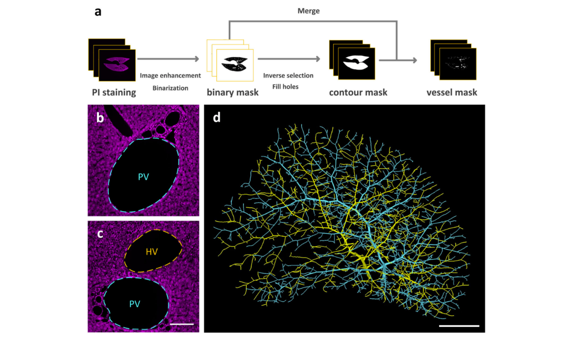
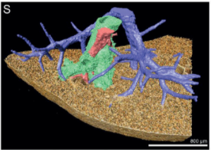
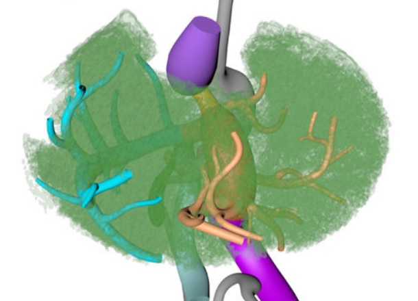
Multiscale reconstruction of various vessels in the intact murine liver lobe
The liver contains a variety of vessels and participates in miscellaneous physiological functions. While past studies generally focused on certain hepatic vessels, we simultaneously obtained all the vessels and cytoarchitectural information of the intact mouse liver lobe at single-cell resolution. […] providing a technology roadmap for studying the fine hepatic vascular structures and their spatial relationship, which will help research into liver diseases and evaluation of medical effi... Read more
Qi Zhang, Anan Li, Siqi Chen, Jing Yuan, Tao Jiang, Xiangning Li, Qingming Luo,Zhao Feng & Hui Gon
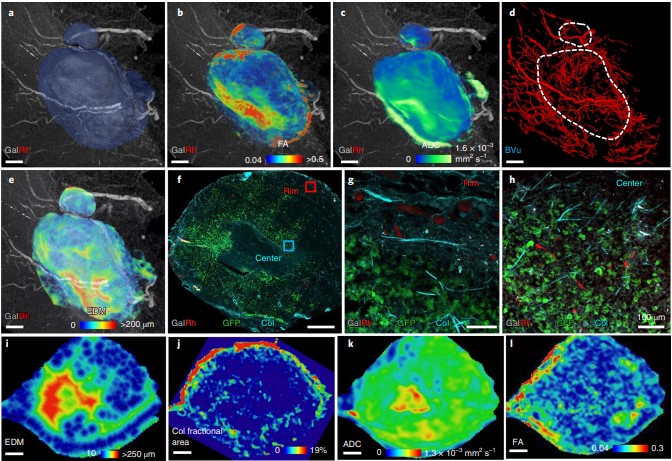
Despite advances in imaging, image-based vascular systems biology has remained challenging because blood vessel data are often available only from a single modality or at a given spatial scale, and cross-modality data are difficult to integrate.
Therefore, there is an exigent need for a multimodality pipeline that enables ex vivo vascular imaging with magnetic resonance imaging, computed tomography and optical microscopy of the same sample, while permitting imaging with complementary c... Read more
Akanksha Bhargava, Benjamin Monteagudo, Priyanka Kushwaha, Janaka Senarathna, Yunke Ren, Ryan C. Riddle , Manisha Aggarwal and Arvind P. Pathak
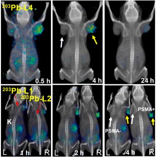
Targeted radiopharmaceutical therapy (TRT) using α-particle radiation is a promising approach for treating both large and micrometastatic lesions. Researchers developed prostate-specific membrane antigen (PSMA)–targeted low-molecular-weight agents for 212Pb-based TRT of patients with prostate cancer (PC) by evaluating the matching γ-emitting surrogate.
Targeted radiopharmaceutical therapy using α-particles (α-TRTs), which cause deposition of ionizing radiation of high-linear-ener... Read more
Sangeeta Ray Banerjee, Il Minn, Vivek Kumar, Anders Josefsson, Ala Lisok, Mary Brummet, Jian Chen, Ana P. Kiess, Kwamena Baidoo, Cory Brayton, Ronnie C. Mease, Martin Brechbiel, George Sgouros, Robert F. Hobbs, and Martin G. Pomper
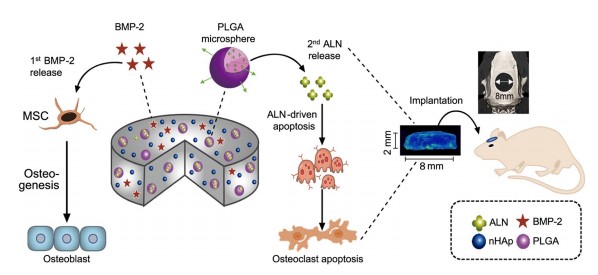
The clinical use of bioactive molecules in bone regeneration has been known to have side effects, which result from uncontrolled and supraphysiological doses.
In this study, we demonstrated the synergistic effect of two bioactive molecules, bone morphogenic protein-2 (BMP-2) and alendronate (ALN), by releasing them in a sequential manner. Collagen-hydroxyapatite composite scaffolds functionalized using BMP-2 are loaded with biodegradable microspheres where ALN is encapsulated.
Th... Read more
Dongtak Lee, Maierdanjiang Wufuer, Insu Kim, Tae Hyun Choi , Byung Jun Kim , Hyo Gi Jung, Byoungjun Jeon, Gyudo Lee, Ok Hee Jeon, Hak Chang & Dae Sung Yoon
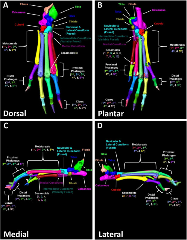
A high-throughput semi-automated bone segmentation workflow for murine hindpaw Micro-CT datasets
Micro-computed tomography (μCT) is a valuable imaging modality for longitudinal quantification of bone volumes to identify disease or treatment effects for a broad range of conditions that affect bone health. Complex structures, such as the hindpaw with up to 31 distinct bones in mice, have considerable analytic potential, but quantification is often limited to a single bone volume metric due to the intensive effort of manual segmentation. Herein, we introduce a high-throughput, user-friendl... Read more
H. Mark Kenney, Yue Peng, Kiana L.Chen, Raquel Ajalik, Lindsay Schnur, Ronald W.Wood, Edward M.Schwarz, Hani A. Awad
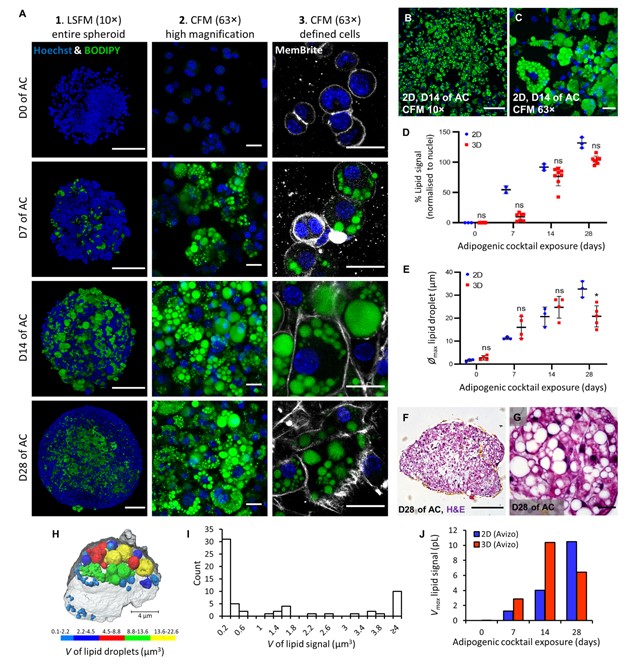
Adipose models have been applied to mechanistic studies of metabolic diseases (such as diabetes) and the subsequent discovery of new therapeutics. However, typical models are either insufficiently complex (2D cell cultures) or expensive and labor intensive (mice/in vivo). To bridge the gap between these models and in order to better inform pre-clinical studies we have developed a drug-responsive 3D model of white adipose tissue (WAT).
Here, spheroids (680 ± 60 μm) comp... Read more
Alexander D Graham, Rajesh Pandey, Viktoriya S Tsancheva, Alessia Candeo, Stanley W Botchway, Alasdair J Allan, Lydia Teboul4, Kamel Madi, Tahkur S Babra, Louisa A K Zolkiewski, Xuan Xue, Liz Bentley, Joan Gannon, Sam N Olof and Roger D Cox
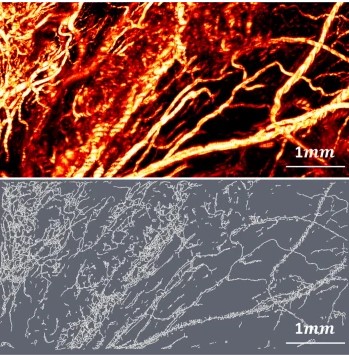
Optical Resolution Photoacoustic Microscopy of Ovary and Fallopian Tube
Ovarian cancer is the leading cause of death among gynecological cancers, but is poorly amenable to preoperative diagnosis. In this study, we investigate the feasibility of “optical biopsy,” using high-optical-resolution photoacoustic microscopy (OR-PAM) to quantify the microvasculature of ovarian and fallopian tube tissue. The technique is demonstrated using excised human ovary and fallopian tube specimens imaged immediately after surgery.
This report describes the first applicatio... Read more
Bin Rao, Xiandong Leng, Yifeng Zeng, Yixiao Lin, Ruimin Chen, Qifa Zhou, Andrea R. Hagemann, Lindsay M. Kuroki, Carolyn K. McCourt, David G. Mutch, Matthew A. Powell, Ian S. Hagemann & Quing Zhu
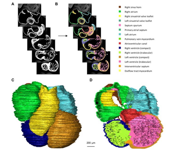
Quantified growth of the human embryonic heart
The size and growth patterns of the components of the human embryonic heart have remained largely undefined.
To provide these data, three-dimensional heart models were generated from immunohistochemically stained sections of ten human embryonic hearts ranging from Carnegie stage 10 to 23. Fifty-eight key structures were annotated and volumetrically assessed. Sizes of the septal foramina and atrioventricular canal opening were also measured. The heart grows exponentially throughout embr... Read more
Jaeike W. Faber, Jaco Hagoort, Antoon F. M. Moorman, Vincent M. Christoffels, Bjarke Jensen
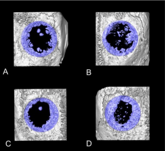
Collagen-Based Matrices for Osteoconduction: A Preclinical In Vivo Study
The aim of this study was to evaluate the influence of additional hydroxyapatite (HA) in collagen-based matrices (CM) and membrane placement on bone formation in calvarial defects.
Critical size defects in the calvaria of 16 New Zealand White Rabbits were randomly treated with CM or mineralized collagen-based matrices (mCM). Half of the sites were covered with a collagen membrane. Animals were euthanized after 12 weeks of healing. The samples were studied by micro-CT and histology. New... Read more
Hiroki Katagiri, Yacine El Tawil, Niklaus P. Lang, Jean-Claude Imber, Anton Sculean, Masako Fujioka-Kobayashi and Nikola Saulacic
Recent treatment developments for metastatic renal cell carcinoma offer combinations of immunotherapies or immunotherapy associated with tyrosine kinase inhibitors (TKI). There is currently no argument to choose one solution or another. Easy-to-use markers to assess longitudinal responses to TKI are necessary to determine when to switch to immunotherapies. These new markers will enable an earlier adaptation of therapeutic strategy in order to prevent tumor development, unnecessary toxicity an... Read more
Alexandre Ingels, Ingrid Leguerney, Paul-Henry Cournède, Jacques Irani, Sophie Ferlicot, Catherine Sébrié, Baya Benatsou, Laurène Jourdain, Stephanie Pitre-Champagnat, Jean-Jacques Patard & Nathalie Lassau
Multiscale Co‐reconstruction of Lung Architectures and Inhalable Materials Spatial Distribution
Pulmonary diseases, such as chronic obstructive pulmonary diseases, lower respiratory infections, and lung cancer are listed in the top ten causes of deaths globally with more than 10 million mortality per year. Apart from oral administration, the Dry Powder Inhalation (DPI) for pulmonary administration provides an important alternative route for targeted treatment of these pulmonary diseases. […] Furthermore, there is a growing demand on und... Read more
Xian Sun, Xiaochuan Zhang, Xiaohong Ren, Hongyu Sun, Li Wu, Caifen Wang, Xiaohui Ye, Peter York, Zhaobing Gao, Hualiang Jiang, Jiwen Zhang, Xianzhen Yin
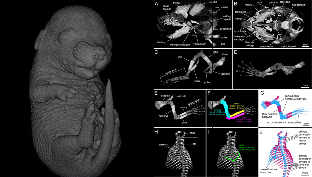
For decades, clearing and staining with Alcian Blue and Alizarin Red has been the gold standard to image vertebrate skeletal development. Here, we present an alternate approach to visualise bone and cartilage based on X-ray microCT imaging, which allows the collection of genuine 3D data of the entire developing skeleton at micron resolution.
Our novel protocol is based on ethanol fixation and staining with Ruthenium Red, and efficiently contrasts cartilage matrix, as demonstrated in wh... Read more
Simone Gabner, Peter Böck, Dieter Fink, Martin Glösmann, Stephan Handschuh
In biomedical research, a huge variety of different techniques is currently available for the structural examination of small specimens, including conventional light microscopy (LM), transmission electron microscopy (TEM), confocal laser scanning microscopy (CLSM), microscopic X-ray computed tomography (microCT), and many others. Since every imaging method is physically limited by certain parameters, a correlative use of complementary methods often yields a significant broader range of inform... Read more
Stephan Handschuh, Natalie Baeumler, Thomas Schwaha & Bernhard Ruthensteiner
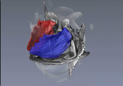
Juvenile Ovine Ex Vivo Larynges: Phonatory, Histologic, and Micro CT Based Anatomic Analyses
It is well known that the phonatory process changes during the life span. However, detailed investigations on potential factors concerned are rare. To deal with this issue, we performed extended biomechanical, macro anatomical, and histological analyses of the contributing laryngeal structures in ex vivo juvenile sheep models. Altogether twelve juvenile sheep larynges were analyzed within the phonatory experiments. Three different elongation levels and 16 different flow levels were applied to... Read more
Michael Döllinger, Olaf Wendler, Claus Gerstenberger, Tanja Grossmann, Marion Semmler, Hossein Sadeghi, and Markus Gugatschka
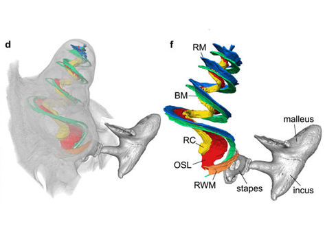
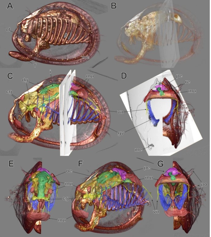
Propagation-based phase-contrast x-ray tomography of cochlea using a compact synchrotron source
We demonstrate that phase retrieval and tomographic imaging at the organ level of small animals can be advantageously carried out using the monochromatic radiation emitted by a compact x-ray light source, without further optical elements apart from source and detector. This approach allows to carry out microtomography experiments which – due to the large performance gap with respect to conventional laboratory instruments – so far were usually limited to synchrotron sources. We dem... Read more
Mareike Töpperwien, Regine Gradl, Daniel Keppeler, Malte Vassholz, Alexander Meyer, Roland Hessler, Klaus Achterhold, Bernhard Gleich, Martin Dierolf, Franz Pfeiffer, Tobias Moser & Tim Salditt
Couinaud based his well-known subdivision of the liver into (surgical) segments on the branching order of portal veins and the location of hepatic veins. However, both segment boundaries and number remain controversial due to an incomplete understanding of the role of liver lobes and vascular physiology on hepatic venous development. Human embryonic livers (5–10 weeks of development) were visualized with Amira 3D-reconstruction and Cinema 4D-remodeling software.
Read more
Jill P. J. M. Hikspoors, Mathijs M. J. P. Peeters, Nutmethee Kruepunga, Hayelom K. Mekonen, Greet M. C. Mommen, S. Eleonore Köhler & Wouter H. Lamers
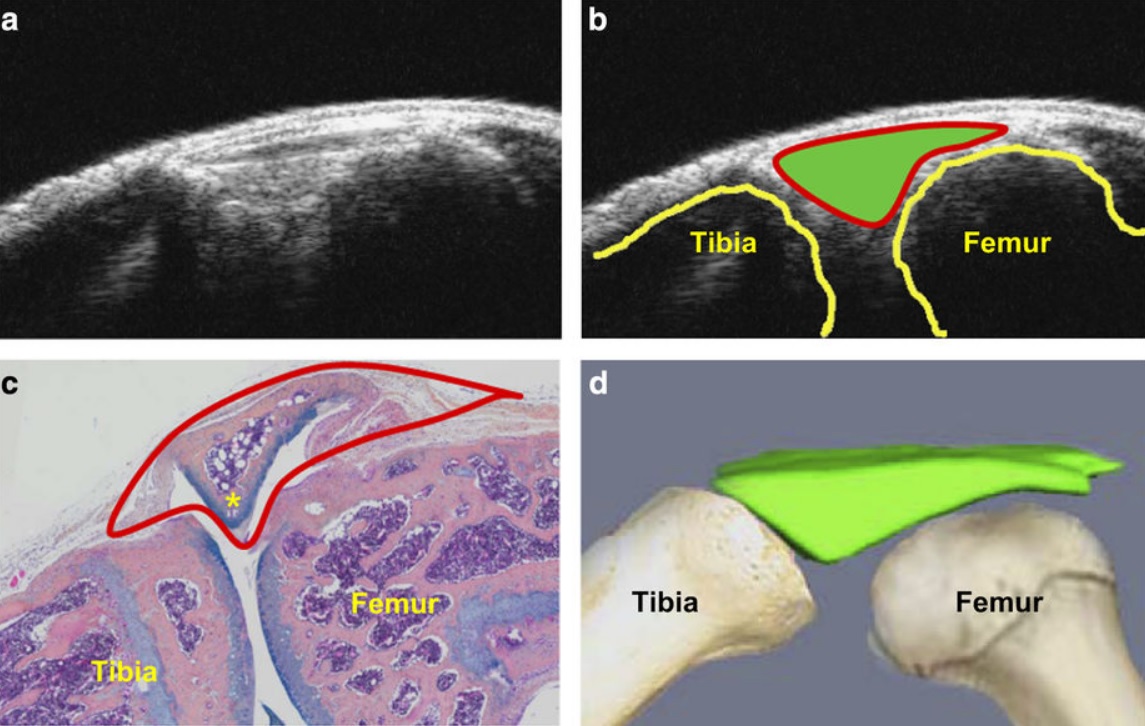
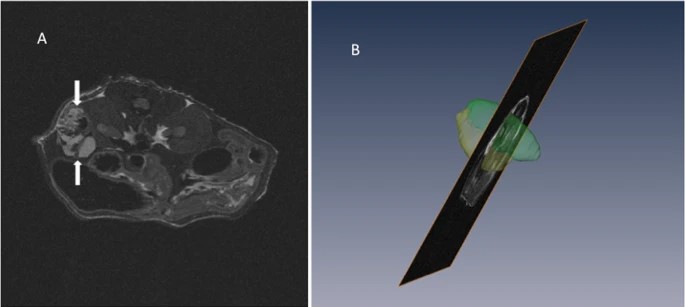
Ultrasound could be a fast and cost-effective means of assessing joint changes in mouse models of posttraumatic osteoarthritis (PTOA). Such models are essential for understanding the biology of this degenerative joint disease and developing new treatments, but noninvasive methods of evaluating disease activity are lacking. Because ultrasound can visualize both joint space volumes and blood flow in the joints, it could provide an alternative to microscopic examination of tissue, assuming it ac... Read more
Hao Xu, Echoe M Bouta, Ronald W Wood, Edward M Schwarz, Yongjun Wang & Lianping Xing